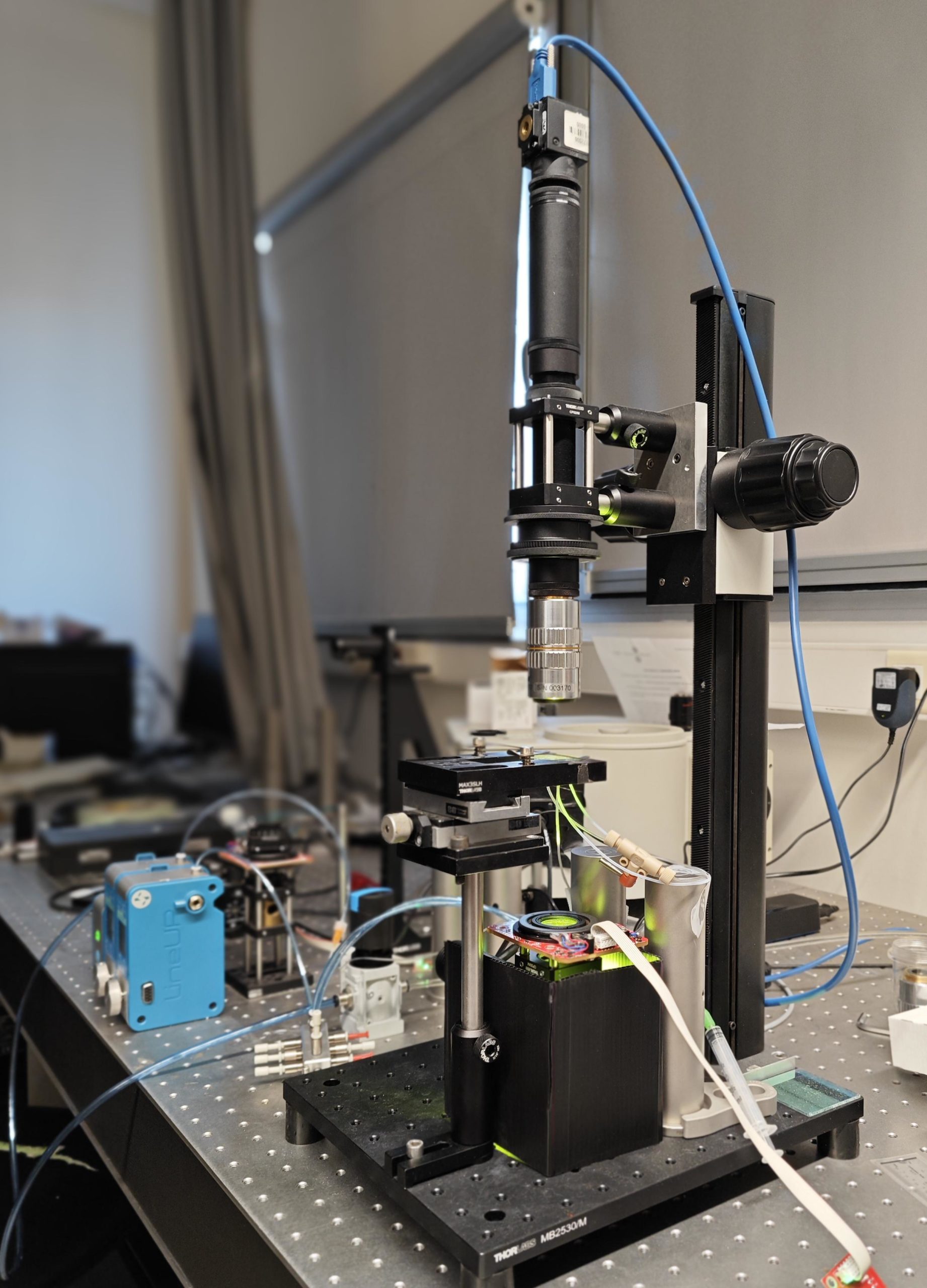
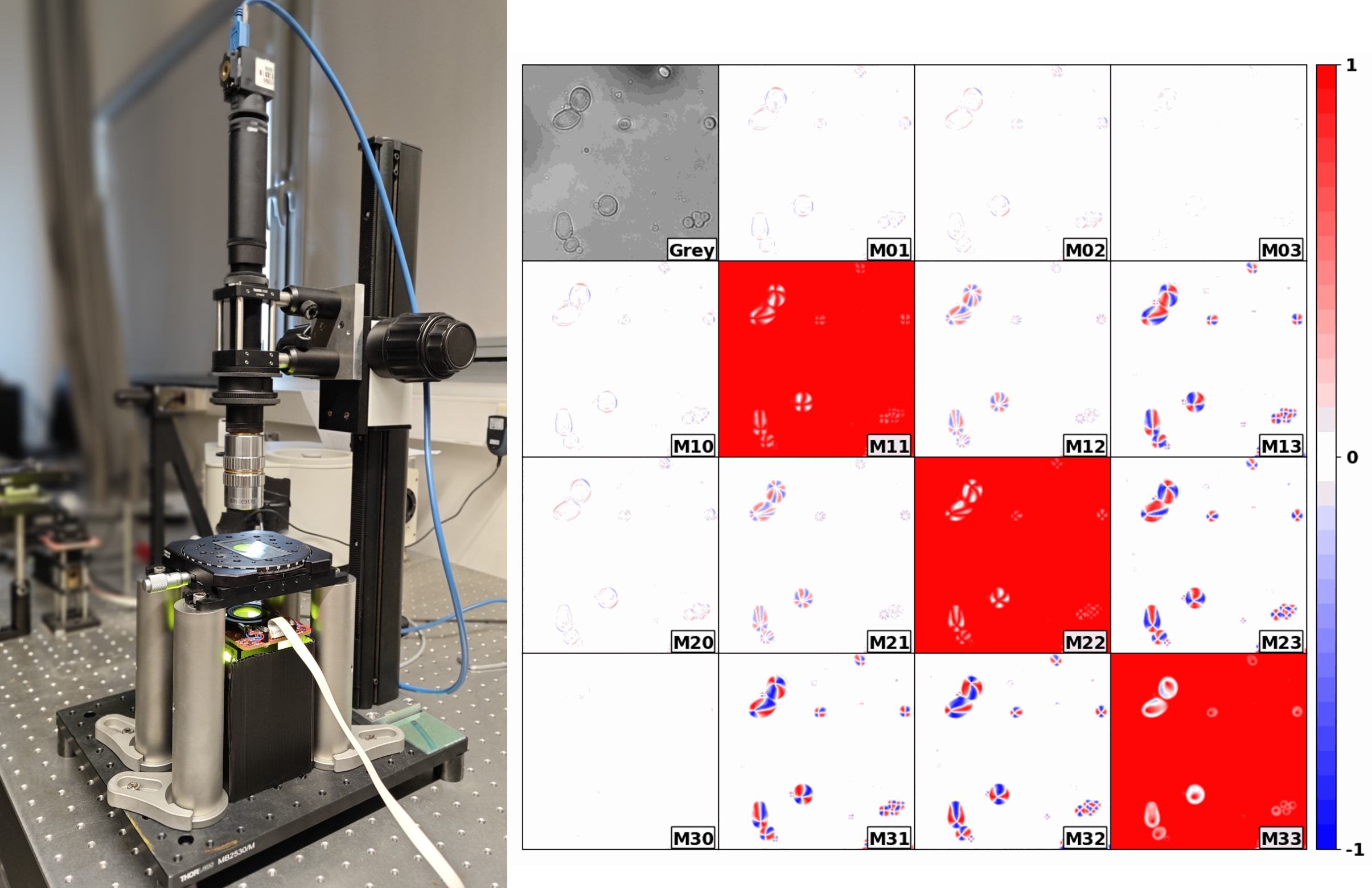
Our Mueller matrix microscope prototype
- High sensitivity and repeatability in distinguishing specific, low-level Mueller Matrix elements.
- Simultaneous measurement of 12 Mueller matrix elements within a rapid timeframe of less than 3 seconds, catering to dynamic applications.
- Real-time measurement capabilities operating at video-rate speed, specifically capturing 2 Mueller Matrix elements for linear birefringence assessments, covering both magnitude and orientation.
- Custom algebraic algorithm to reconstruct the full 16 MM elements from the 12 measured MM elements (in the absence of depolarization)
- Adjustable to different wavelengths (VIS and NIR)
- Software packages with data analysis features for all 6 polarization parameters (linear retardation – magnitude and angle; circular retardation – magnitude; linear diattenuation – magnitude and angle; and circular diattenuation – magnitude).
- Optimal optical and polarimetric design
- engineered for cost efficiency without compromising on performance.
- Highly customizable layout.
How it works?
The system employs a rotating compensator in the polarization state generator (PSG) and a polarization camera as detector. Importantly, no moving parts are present between the sample and the objective, preserving image stability and optical alignment. The rotating compensator operates in step-scan mode, decoupling mechanical rotation from image acquisition and allowing for independent control of camera exposure.
Are results quantative?
Yes. Every pixel stores a 12-element Mueller matrix, which can be post-processed using our analytical inversion algorithm to retrieve the full set of polarization parameters mentioned above.
What are the advantages and disadvantages of this technology with respect to other technologies?
The main competing systems typically use liquid crystal modulators or photoelastic modulators (PEMs).
Liquid Crystal Modulators (LCMs)
-
Pros: Fast switching, no mechanical motion.
-
Cons: Require thermal stabilization, frequent recalibrations, and suffer from non-uniformity across the aperture.
Photoelastic Modulators (PEMs)
-
Pros: Excellent for high-speed spectroscopy.
-
Cons: Incompatible with standard video-rate cameras. Strobing techniques reduce light throughput. The high cost of PEMs is not justified unless used with spectrometers, not cameras.
Our system avoids these limitations. It provides stable, high-throughput, and quantitative measurements, while being cost-efficient and easily customizable
What spatial resolutions are possible? Are there multiple versions?
Yes, we currently operate two distinct implementations:
-
A microscope version with objectives ranging from 2× to 50×, achieving spatial resolutions from a few millimeters down to a few micrometers.
-
A wide-field imaging version suited for larger fields of view, with centimeter-scale spatial resolution, ideal for applications like imaging entire histological slides.
How is it calibrated?
Calibration is fully automated and requires no calibration samples. It is performed with a simple click, using either an empty stage or a blank glass sample. Once calibrated, the system remains stable over time. Recalibration is only necessary if the LED source is changed or the camera alignment is modified.
Can do real time imaging?
Yes, with the “live mode” the instrument can image at 50fps the azimuth and magnitude of linear retardation of transparent samples. For more complex samples that require more information (linear diattenuation, circular retardaction, circular diattenuation) the Mueller matrix measurement takes 3 seconds. The system alows for continous measurement of Mueller matrices and saving.
Is the limited extinction ratio of the polarization camera a limitation?
No, it is not a limitation in our system. The Sony Polarsens™ sensor used in our polarization camera offers pixel-level extinction ratios better than 1:100, which is sufficient for accurate and quantitative Mueller matrix measurements. The extinction is also uniform across the field, unlike liquid crystals or other modulators. In practice, the camera’s performance fully supports the precision and sensitivity required by our applications. Only above 750nm-800nm camera’s polarization performance can be a bootleneck for measurement quality.
Is it limited to one wavelength?
Not at all. The system can be adjusted across visible and near-infrared wavelengths (VIS–NIR). Changing wavelength is as simple as replacing the illumination LED and one click calibration.We’ve successfully operated the microscope across the 405–980 nm range, with best performance typically around 550 nm. For full spectral analysis, we can also couple the system with a monochromator and a halogen source, though this is most practical at lower magnifications due to light throughput considerations.
What are the applications?
Over the past months and years, we’ve applied this system to a variety of scientific challenges. For example it has been used to:
-
Identify chiral particles in complex mixtures
-
Analyze collagen fiber orientation in heart tissue
-
Perform stress mapping in transparent solids
-
Characterize histological sections
-
Monitor crystal growth in microfluidic devices
What software do you use?
All our software have been self-developed in Labview.
Examples
Film of biofilaments from cytoskeleton proteins. Self-propelled system.
Intensity imaging (what a normal microscope can see)
Live-birefrigence magnitude imaging (what our microscope can measure in real-time)
We usually report birefringence values in radians but they can be easily converted to nm. In this case, the 0.08 radians shown in the scale are equivalent to 7 nm of birefringence: 0.08*550 nm/(2*pi).
Full MM in a growing crystal
The optical layout is very flexible to accommodate different different types of in-situ experiments. This is a photo of our prototype doing measurements in microfluidic chambers